


Let us help you find what you're looking for.
13 May 2025
.jpg)
WHAT IS DXA FFI?
WHAT ARE ATYPICAL FEMORAL FRACTURES?
KEY BENEFITS OF DXA FFI

EARLY DETECTION'S CRITICAL ROLE: A CASE EXAMPLE

KEY TAKEAWAYS FOR GPs
WHO AND HOW GPs CAN REFER
WHAT IS DXA FFI?
Dual-energy X-ray absorptiometry (DXA) full-length femur imaging (FFI) to improve the early detection of atypical femoral fractures (AFFs) is now offered at the Department of Nuclear Medicine and Molecular Imaging at Singapore General Hospital (SGH).
This new imaging technique is tailored for at-risk patients, particularly those receiving long-term bisphosphonate or denosumab therapy.
DXA FFI employs low-dose radiation to provide detailed visualisation of the entire femur, aiding the detection of precursors to AFFs, including cortical thickening and stress fractures.
WHAT ARE ATYPICAL FEMORAL FRACTURES?
AFFs are stress or insufficiency fractures typically occurring in the femoral shaft, associated with long-term bisphosphonate or denosumab use. These fractures can occur without trauma and result from suppressed bone remodelling, microdamage accumulation, and mechanical factors.
Higher-risk groups include Asian patients, those on glucocorticoids, and individuals with prolonged bisphosphonate/denosumab use (≥3 years).
WHEN DXA FFI IS RECOMMENDED
The International Society for Clinical Densitometry (ISCD) recommends FFI for patients currently on or who have recently discontinued bisphosphonate or denosumab therapy (within one year), particularly those with ≥ 3 years of cumulative exposure or concurrent glucocorticoid use.
This screening tool is suitable for patients with no prodromal symptoms such as pain.
KEY BENEFITS OF DXA FFI
- Early detection: Identifies changes before complete fractures occur, allowing timely intervention
- Safe and non-invasive nature: Quick (under five minutes per femur), painless and with low radiation exposure – making it ideal for regular monitoring
- Comprehensive assessment: Bilateral imaging enables detailed comparisons, improving diagnostic accuracy
- Convenience: Performed alongside routine bone mineral density (BMD) assessment
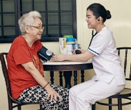
- Patient-friendliness: Minimal preparation required, with optimised positioning aids for patient comfort (Figure 1)
Figure 1 Nuclear Medicine technologist positioning a patient for FFI, using dedicated positioning aids to ensure proper alignment and patient comfort during the procedure at the Department of Nuclear Medicine and Molecular Imaging, Singapore General Hospital (SGH).
HOW GPs CAN MANAGE AT-RISK PATIENTS UNSUITABLE FOR DXA FFI
Patients with prodromal symptoms are not suitable for DXA FFI.
For at-risk patients presenting with thigh, hip or groin pain:
- Confirm diagnosis through plain radiography
- Consider temporary discontinuation of bisphosphonates/denosumab
- Limit weight-bearing activities and prescribe crutches if necessary
- Refer to orthopaedics upon AFF confirmation
EARLY DETECTION'S CRITICAL ROLE: A CASE EXAMPLE
A post-menopausal patient with osteoporosis, on alendronate for over five years, underwent FDG PET/CT scanning for left lower lobe adenocarcinoma staging. Incidental findings revealed cortical thickening in the bilateral femoral mid-shafts, suggesting incomplete AFFs.
Plain radiographs confirmed these incomplete fractures (Figure 2). Three days later, the patient sustained a displaced left proximal femoral shaft fracture following minor trauma, necessitating intramedullary nailing and extended rehabilitation.
This case emphasises the vital importance of early detection and intervention in preventing severe outcomes.
Figure 2 18F-FDG PET/CT images demonstrating cortical thickening in the bilateral femoral mid-shafts (arrows). These findings were corroborated by conventional radiographic imaging.
KEY TAKEAWAYS FOR GPs
- DXA FFI can now be routinely incorporated with BMD assessment for at-risk patients.
- GPs play a crucial role in identifying at-risk patients and recommending appropriate screening to prevent complications.
WHO AND HOW GPs CAN REFER
GPs can refer patients via the SGH GP Network or visit the website for more information on the procedure.
Tel: 6326 6060
Email: gpnetwork@sgh.com.sg

Stay Healthy The Easy Way
Get trusted health advice, offers and more.
Stay Ahead in Healthcare Industry
Subscribe to our exclusive updates for healthcare professionals.


Stay Healthy With
Follow SGH


